

Radiation-based imaging technique.

An ascending cystourethrogram is a type of X-ray that is used to examine the urethra, which is the tube that carries urine from the bladder to the outside of the body. During this procedure, a contrast is injected into the urethra through the tip of the penis, and X-rays are taken to visualize the flow of the contrast. This test is generally carried out in males and is also known as ascending urethrography.
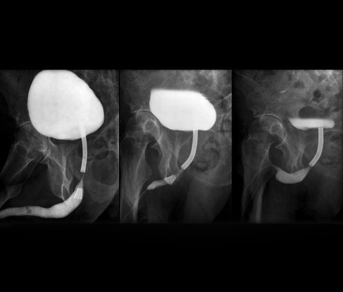
An Ascending Cystourethrogram (VCUG) is a diagnostic imaging procedure that involves the injection of contrast dye into the bladder through a catheter, followed by X-ray imaging to evaluate the anatomy and function of the bladder and lower urinary tract. Here are some common reasons why a healthcare provider might recommend a VCUG:
Recurrent Urinary Tract Infections (UTIs): VCUG is often used to assess for abnormalities in the urinary tract that may contribute to recurrent UTIs, especially in children.
Vesicoureteral Reflux (VUR): VCUG is a primary diagnostic tool for evaluating vesicoureteral reflux, a condition where urine flows backward from the bladder into the ureters and possibly into the kidneys. VUR can increase the risk of UTIs and kidney damage.
Congenital Anomalies: It is used to detect congenital abnormalities in the structure of the urinary tract, such as urethral strictures, bladder diverticula, or abnormalities in the ureterovesical junction.
Urinary Incontinence: VCUG can be part of the evaluation for urinary incontinence, helping to identify issues such as bladder dysfunction or structural abnormalities.
Voiding Dysfunction: It is employed to assess voiding dysfunction or difficulties in emptying the bladder, which may be related to anatomical or functional issues.
Bladder Outlet Obstruction: VCUG can help identify conditions that cause obstruction at the bladder outlet, such as urethral strictures or enlarged prostate in males.
Monitoring Post-Surgical Procedures: In some cases, VCUG is used to monitor the results of surgical interventions on the urinary tract, such as repairs to correct structural abnormalities.
Evaluation of Neurogenic Bladder: VCUG may be recommended for individuals with suspected neurogenic bladder dysfunction, which can result from conditions affecting the nervous system.
It’s important to note that VCUG involves the use of radiation and contrast dye, and the decision to undergo this procedure is based on a careful assessment of the individual’s medical history, symptoms, and the information needed for diagnosis. Alternative imaging modalities, such as ultrasound or magnetic resonance imaging (MRI), may be considered depending on the specific clinical situation.
The preparation for a spine X-ray is generally straightforward, and there are usually minimal requirements. However, it’s essential to follow any specific instructions given by your healthcare provider or the imaging facility. Here are some general guidelines:
Clothing: You may be asked to change into a hospital gown, as buttons, zippers, or other metal components in regular clothing can interfere with the X-ray images.
Remove Metal Objects: You’ll likely be asked to remove any metal objects or jewelry from the area being X-rayed, as metal can interfere with the quality of the images. This includes items such as belts, watches, necklaces, piercings, and even underwire bras.
Inform the Technologist: Make sure to inform the X-ray technologist if there is any chance you could be pregnant, as they may need to take additional precautions to minimize radiation exposure to the fetus.
Follow Instructions for Positioning: The technologist will guide you into the appropriate positions for the X-ray. It’s important to follow their instructions to ensure accurate and clear images.
Stay Still during the Procedure: To obtain clear images, it’s crucial to remain still while the X-rays are being taken. The technologist may use immobilization devices or props to help you maintain the required positions.
Discuss Medical History: Inform the technologist and your healthcare provider about any relevant medical history, including prior surgeries or conditions that might affect the spine. This information helps ensure the imaging is tailored to your specific needs.
Generally, X-rays are safe and relatively quick procedures. The amount of radiation exposure is kept as low as reasonably achievable while still obtaining the necessary diagnostic information. If you have any concerns or questions about the procedure or its potential risks, don’t hesitate to discuss them with your healthcare provider or the radiology staff.
