

Radiation-based imaging technique.

Barium tests are used to help see the outline of various parts of the gut (gastrointestinal tract). These include the oesophagus, stomach, small intestines and colon.
Barium X-ray tests are done less commonly these days. Today we usually look into the gut with a flexible endoscopy or colonoscopy. However, there is still a place for barium tests to help assess various problems of the gut.
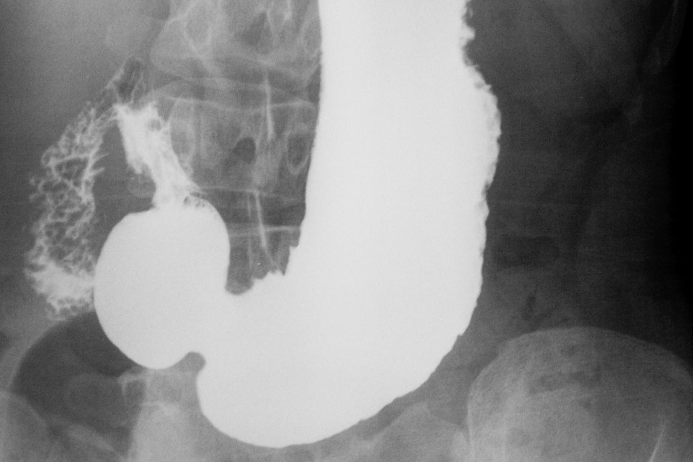
A Barium Meal and Swallow, also known as an upper gastrointestinal (GI) series or barium swallow, is a diagnostic imaging test that involves the use of barium contrast material to visualize the upper part of the digestive system, including the esophagus, stomach, and small intestine. Here are some common reasons why a healthcare provider might recommend a Barium Meal and Swallow:
Swallowing Difficulties (Dysphagia): The test is often used to evaluate difficulties with swallowing, which may be caused by conditions such as esophageal strictures, tumors, or motility disorders.
Gastroesophageal Reflux Disease (GERD): It can be employed to assess for signs of GERD, a condition where stomach acid flows back into the esophagus, causing symptoms such as heartburn and regurgitation.
Hiatal Hernia: Barium swallow can help in diagnosing hiatal hernias, where part of the stomach protrudes through the diaphragm into the chest cavity.
Esophageal Tumors or Lesions: The test can detect the presence of tumors, ulcers, or other abnormal growths in the esophagus.
Stomach Ulcers: Barium meal and swallow can visualize the stomach lining and identify ulcers or erosions.
Malabsorption Disorders: It may be used to assess the small intestine for abnormalities that could contribute to malabsorption disorders.
Crohn’s Disease: In some cases, the test may be used to evaluate the presence and extent of Crohn’s disease affecting the upper gastrointestinal tract.
Preoperative Planning: Barium swallow may be performed before certain surgical procedures on the digestive system to provide detailed information about the anatomy and potential issues.
The procedure involves drinking a barium solution, which is a chalky liquid that coats the lining of the digestive tract and makes it visible on X-ray images. X-rays are then taken as the contrast material moves through the esophagus and into the stomach and small intestine.
The preparation for a spine X-ray is generally straightforward, and there are usually minimal requirements. However, it’s essential to follow any specific instructions given by your healthcare provider or the imaging facility. Here are some general guidelines:
Clothing: You may be asked to change into a hospital gown, as buttons, zippers, or other metal components in regular clothing can interfere with the X-ray images.
Remove Metal Objects: You’ll likely be asked to remove any metal objects or jewelry from the area being X-rayed, as metal can interfere with the quality of the images. This includes items such as belts, watches, necklaces, piercings, and even underwire bras.
Inform the Technologist: Make sure to inform the X-ray technologist if there is any chance you could be pregnant, as they may need to take additional precautions to minimize radiation exposure to the fetus.
Follow Instructions for Positioning: The technologist will guide you into the appropriate positions for the X-ray. It’s important to follow their instructions to ensure accurate and clear images.
Stay Still during the Procedure: To obtain clear images, it’s crucial to remain still while the X-rays are being taken. The technologist may use immobilization devices or props to help you maintain the required positions.
Discuss Medical History: Inform the technologist and your healthcare provider about any relevant medical history, including prior surgeries or conditions that might affect the spine. This information helps ensure the imaging is tailored to your specific needs.
Generally, X-rays are safe and relatively quick procedures. The amount of radiation exposure is kept as low as reasonably achievable while still obtaining the necessary diagnostic information. If you have any concerns or questions about the procedure or its potential risks, don’t hesitate to discuss them with your healthcare provider or the radiology staff.
