

Radiation-based imaging technique.

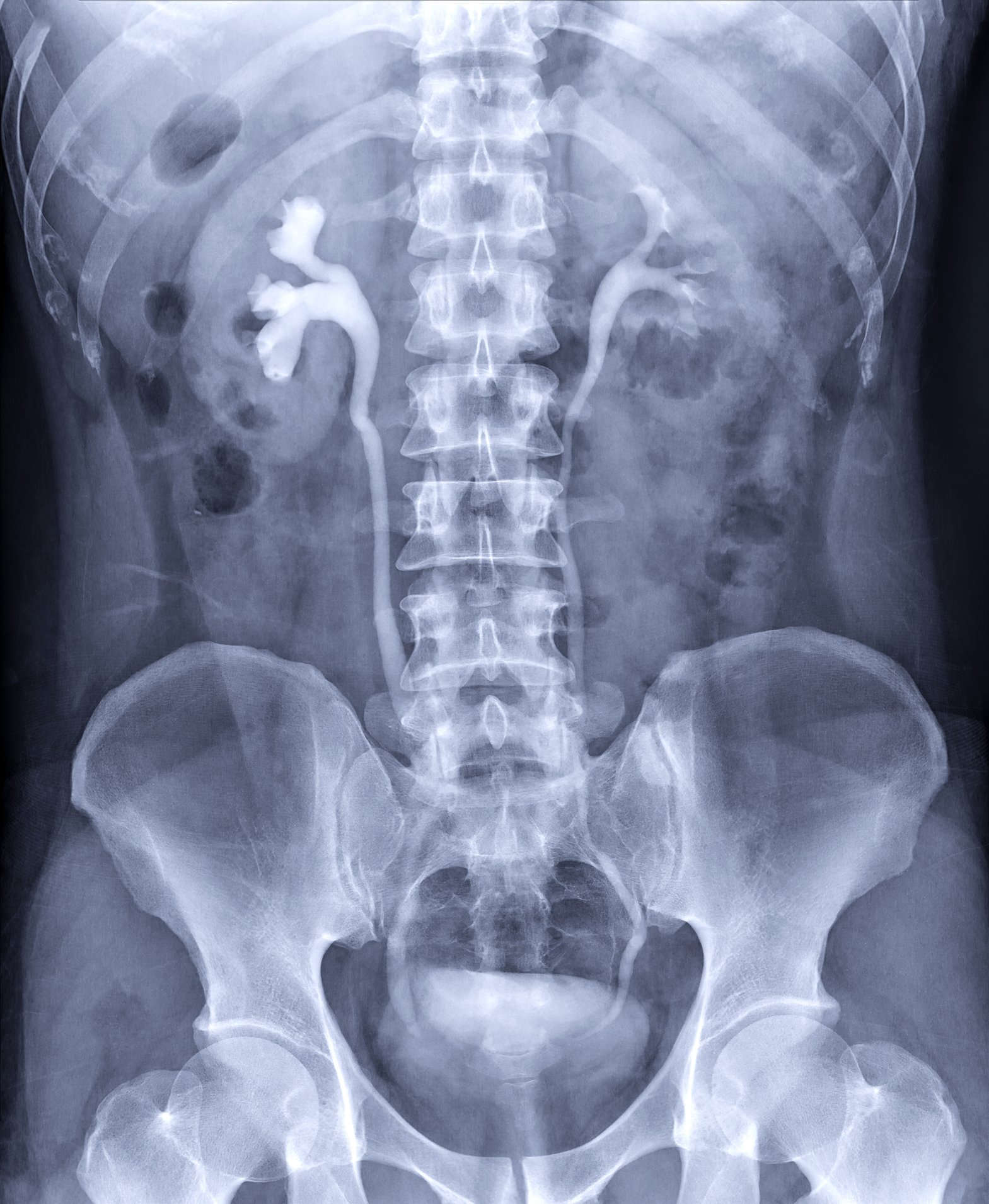
Intravenous Pyelogram (IVP) is an x-ray exam that uses a special dye to outline the kidneys, ureters and bladder. It can show how your renal and urinary system handles fluid waste. This helps your health care team find problems in the urinary tract.
An IVP may provide enough information to allow your doctor to treat you with medication and avoid surgery.
The procedure involves injecting a contrast material into a vein in the patient’s arm, which travels through the blood stream and collects in the kidneys and urinary tract, turning these areas bright white on the x-ray images.
The exam is used to help diagnose symptoms such as blood in the urine or pain in the side or lower back.
If you are scheduled for an IVP, your doctor will give you detailed instructions on how to prepare for the exam. You will likely be instructed not to eat or drink after midnight on the night before your exam. You may also be asked to take a mild laxative (in either pill or liquid form) the evening before the procedure. Inform your doctor if there’s a possibility you are pregnant and discuss any recent illnesses, medical conditions, medications you’re taking and allergies, especially to iodine-based contrast materials.
An Intravenous Pyelogram (IVP) is a radiological procedure used to examine the urinary system, including the kidneys, ureters, and bladder. It involves the injection of a contrast dye into a vein, usually in the arm, followed by a series of X-rays to capture images of the urinary tract. Here are some common reasons why a healthcare provider might recommend an IVP:
Kidney Stones: IVP is often used to detect the presence of kidney stones and assess their size, location, and the degree of obstruction they may be causing in the urinary system.
Urinary Tract Obstruction: It helps in identifying and locating any blockages or obstructions in the urinary tract, such as those caused by tumors, strictures, or congenital abnormalities.
Hematuria (Blood in Urine): IVP can be used to investigate the cause of hematuria, which is the presence of blood in the urine. It helps in identifying the source of bleeding within the urinary system.
Structural Abnormalities: IVP is useful for detecting and evaluating structural abnormalities in the kidneys, ureters, and bladder, including congenital anomalies or acquired conditions.
Renal Function Assessment: While not a direct measure of renal function, IVP can provide indirect information about kidney function by assessing the excretion of contrast material.
Urinary Tract Infections (UTIs): It may be used in certain cases to evaluate the impact of urinary tract infections on the structures of the urinary system.
Preoperative Planning: Prior to certain surgical procedures involving the urinary system, an IVP may be performed to provide detailed information about the anatomy and any existing abnormalities.
Follow-up of Previous Conditions: IVP may be used for follow-up assessments after certain treatments or surgeries to monitor changes in the urinary system.
It’s important to note that while IVP has been a common diagnostic tool, other imaging modalities such as computed tomography (CT) urography and magnetic resonance urography (MRU) are increasingly used for similar purposes and may provide more detailed and three-dimensional images.
The preparation for a spine X-ray is generally straightforward, and there are usually minimal requirements. However, it’s essential to follow any specific instructions given by your healthcare provider or the imaging facility. Here are some general guidelines:
Clothing: You may be asked to change into a hospital gown, as buttons, zippers, or other metal components in regular clothing can interfere with the X-ray images.
Remove Metal Objects: You’ll likely be asked to remove any metal objects or jewelry from the area being X-rayed, as metal can interfere with the quality of the images. This includes items such as belts, watches, necklaces, piercings, and even underwire bras.
Inform the Technologist: Make sure to inform the X-ray technologist if there is any chance you could be pregnant, as they may need to take additional precautions to minimize radiation exposure to the fetus.
Follow Instructions for Positioning: The technologist will guide you into the appropriate positions for the X-ray. It’s important to follow their instructions to ensure accurate and clear images.
Stay Still during the Procedure: To obtain clear images, it’s crucial to remain still while the X-rays are being taken. The technologist may use immobilization devices or props to help you maintain the required positions.
Discuss Medical History: Inform the technologist and your healthcare provider about any relevant medical history, including prior surgeries or conditions that might affect the spine. This information helps ensure the imaging is tailored to your specific needs.
Generally, X-rays are safe and relatively quick procedures. The amount of radiation exposure is kept as low as reasonably achievable while still obtaining the necessary diagnostic information. If you have any concerns or questions about the procedure or its potential risks, don’t hesitate to discuss them with your healthcare provider or the radiology staff.
