

Radiation-based imaging technique.

The adenoid is a mass of lymphatic tissue located behind the nasal cavity, in the roof of the nasopharynx, where the nose blends into the throat. The adenoid, unlike the palatine tonsils, has pseudostratified epithelium. The adenoids are part of the so-called Waldeyer ring of lymphoid tissue which also includes the palatine tonsils, the lingual tonsils and the tubal tonsils
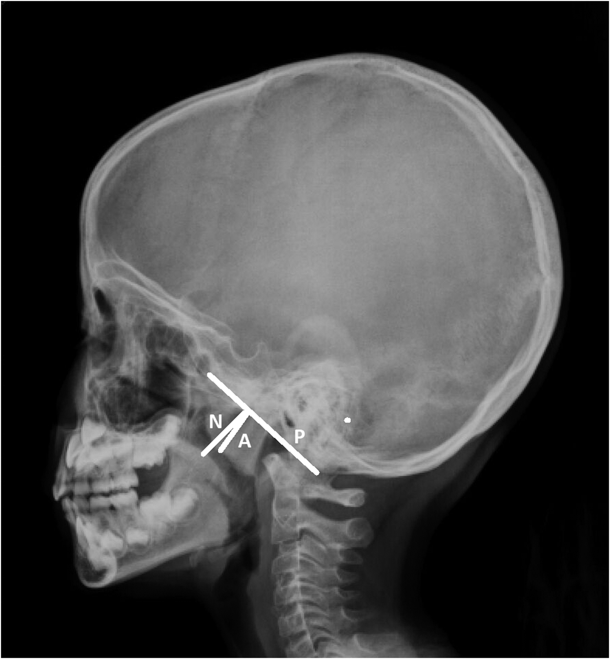
X-rays of the adenoids may be performed for specific medical reasons to assess and diagnose conditions related to the adenoid tissue, which is a part of the lymphoid tissue in the back of the nasal cavity. Here are some common indications for getting an X-ray of the adenoids:
Enlarged Adenoids: X-rays can be used to evaluate the size of the adenoids. Enlarged adenoids can lead to various symptoms, such as nasal congestion, difficulty breathing, snoring, or recurrent ear infections.
Chronic Sinus Infections: Adenoid X-rays may be part of the diagnostic process for individuals with chronic or recurrent sinus infections to determine if enlarged adenoids are contributing to the problem.
Ear Infections: Enlarged adenoids can block the Eustachian tubes, leading to ear infections. X-rays may be used to assess the size of the adenoids and their potential impact on the Eustachian tubes.
Sleep Apnea: Adenoid X-rays may be considered in cases where sleep apnea is suspected, especially in children. Enlarged adenoids can obstruct the airway during sleep, contributing to breathing difficulties.
Nasal Obstruction: X-rays can help assess the extent to which enlarged adenoids may be causing nasal obstruction or congestion.
It’s important to note that while X-rays can provide information about the size and structure of the adenoids, they may not provide detailed information about the functional aspects of the adenoid tissue. In some cases, other imaging modalities or diagnostic tools, such as endoscopy, may be used for a more comprehensive evaluation.
The preparation for a spine X-ray is generally straightforward, and there are usually minimal requirements. However, it’s essential to follow any specific instructions given by your healthcare provider or the imaging facility. Here are some general guidelines:
Clothing: You may be asked to change into a hospital gown, as buttons, zippers, or other metal components in regular clothing can interfere with the X-ray images.
Remove Metal Objects: You’ll likely be asked to remove any metal objects or jewelry from the area being X-rayed, as metal can interfere with the quality of the images. This includes items such as belts, watches, necklaces, piercings, and even underwire bras.
Inform the Technologist: Make sure to inform the X-ray technologist if there is any chance you could be pregnant, as they may need to take additional precautions to minimize radiation exposure to the fetus.
Follow Instructions for Positioning: The technologist will guide you into the appropriate positions for the X-ray. It’s important to follow their instructions to ensure accurate and clear images.
Stay Still during the Procedure: To obtain clear images, it’s crucial to remain still while the X-rays are being taken. The technologist may use immobilization devices or props to help you maintain the required positions.
Discuss Medical History: Inform the technologist and your healthcare provider about any relevant medical history, including prior surgeries or conditions that might affect the spine. This information helps ensure the imaging is tailored to your specific needs.
Generally, X-rays are safe and relatively quick procedures. The amount of radiation exposure is kept as low as reasonably achievable while still obtaining the necessary diagnostic information. If you have any concerns or questions about the procedure or its potential risks, don’t hesitate to discuss them with your healthcare provider or the radiology staff.
