

Radiation-based imaging technique.

A spinal X-ray is a procedure that uses radiation to make detailed pictures of the bones of your spine. It can help your doctor find out what’s causing your back or neck pain.
A technician uses a machine that sends X-ray beams through your body. It records a black-and-white image on a special film or computer. Bones, and other parts of your body that are thick or dense, show up white in the picture. Softer tissue, like fat or muscle, appears in shades of gray.
Your doctor can take separate X-rays that focus on the different parts of the spine, which is made up of 33 small bones called vertebrae.
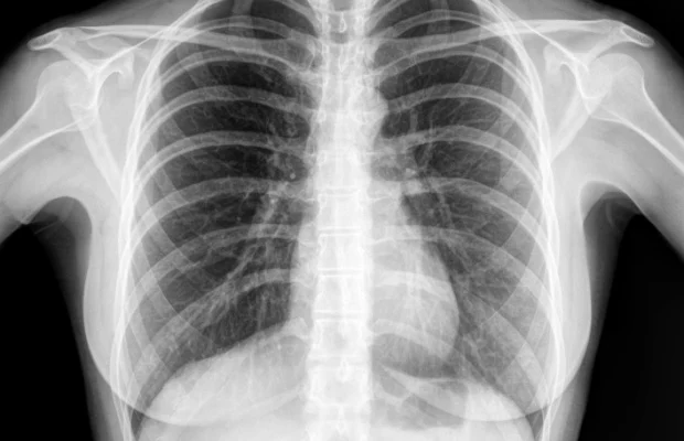
X-rays of the chest, often referred to as chest X-rays, are commonly performed to assess the structures within the chest cavity, including the heart and lungs. Here are some reasons why a healthcare provider might recommend a chest X-ray to evaluate the heart:
Cardiopulmonary Conditions: Chest X-rays are often used to evaluate and diagnose various cardiopulmonary conditions, including heart and lung diseases. They can provide information about the size, shape, and position of the heart and its major vessels.
Heart Enlargement: An X-ray can help identify if the heart is enlarged, which may be indicative of conditions such as cardiomyopathy, heart failure, or other cardiac disorders.
Congestive Heart Failure: Chest X-rays may be used to assess signs of congestive heart failure, such as fluid accumulation in the lungs (pulmonary edema).
Chest Pain or Discomfort: If you are experiencing chest pain or discomfort, a chest X-ray may be ordered to rule out or identify potential cardiac causes, such as coronary artery disease or other heart-related issues.
Pulmonary Conditions: Chest X-rays are valuable for diagnosing and monitoring lung conditions that may affect the heart, such as pneumonia, chronic obstructive pulmonary disease (COPD), or lung cancer.
Trauma: Following chest trauma, X-rays may be used to assess for injuries to the chest wall, ribs, or other structures, including the heart.
Preoperative Assessment: Prior to certain cardiac or thoracic surgeries, chest X-rays may be performed as part of the preoperative assessment to provide baseline information about the heart and lungs.
It’s important to note that while chest X-rays can provide valuable information about the heart and lungs, they have limitations in terms of visualizing fine details and may not be as comprehensive as other imaging modalities such as echocardiography, CT scans, or MRI. The choice of imaging modality depends on the specific clinical situation and the information needed by the healthcare provider.
The preparation for a spine X-ray is generally straightforward, and there are usually minimal requirements. However, it’s essential to follow any specific instructions given by your healthcare provider or the imaging facility. Here are some general guidelines:
Clothing: You may be asked to change into a hospital gown, as buttons, zippers, or other metal components in regular clothing can interfere with the X-ray images.
Remove Metal Objects: You’ll likely be asked to remove any metal objects or jewelry from the area being X-rayed, as metal can interfere with the quality of the images. This includes items such as belts, watches, necklaces, piercings, and even underwire bras.
Inform the Technologist: Make sure to inform the X-ray technologist if there is any chance you could be pregnant, as they may need to take additional precautions to minimize radiation exposure to the fetus.
Follow Instructions for Positioning: The technologist will guide you into the appropriate positions for the X-ray. It’s important to follow their instructions to ensure accurate and clear images.
Stay Still during the Procedure: To obtain clear images, it’s crucial to remain still while the X-rays are being taken. The technologist may use immobilization devices or props to help you maintain the required positions.
Discuss Medical History: Inform the technologist and your healthcare provider about any relevant medical history, including prior surgeries or conditions that might affect the spine. This information helps ensure the imaging is tailored to your specific needs.
Generally, X-rays are safe and relatively quick procedures. The amount of radiation exposure is kept as low as reasonably achievable while still obtaining the necessary diagnostic information. If you have any concerns or questions about the procedure or its potential risks, don’t hesitate to discuss them with your healthcare provider or the radiology staff.
