

Radiation-based imaging technique.

A skull X-ray is a series of pictures of the bones of the skull. Skull X-rays have largely been replaced by computed tomography (CT) scans. A skull X-ray may help find head injuries, bone fractures, or abnormal growths or changes in bone structure or size.
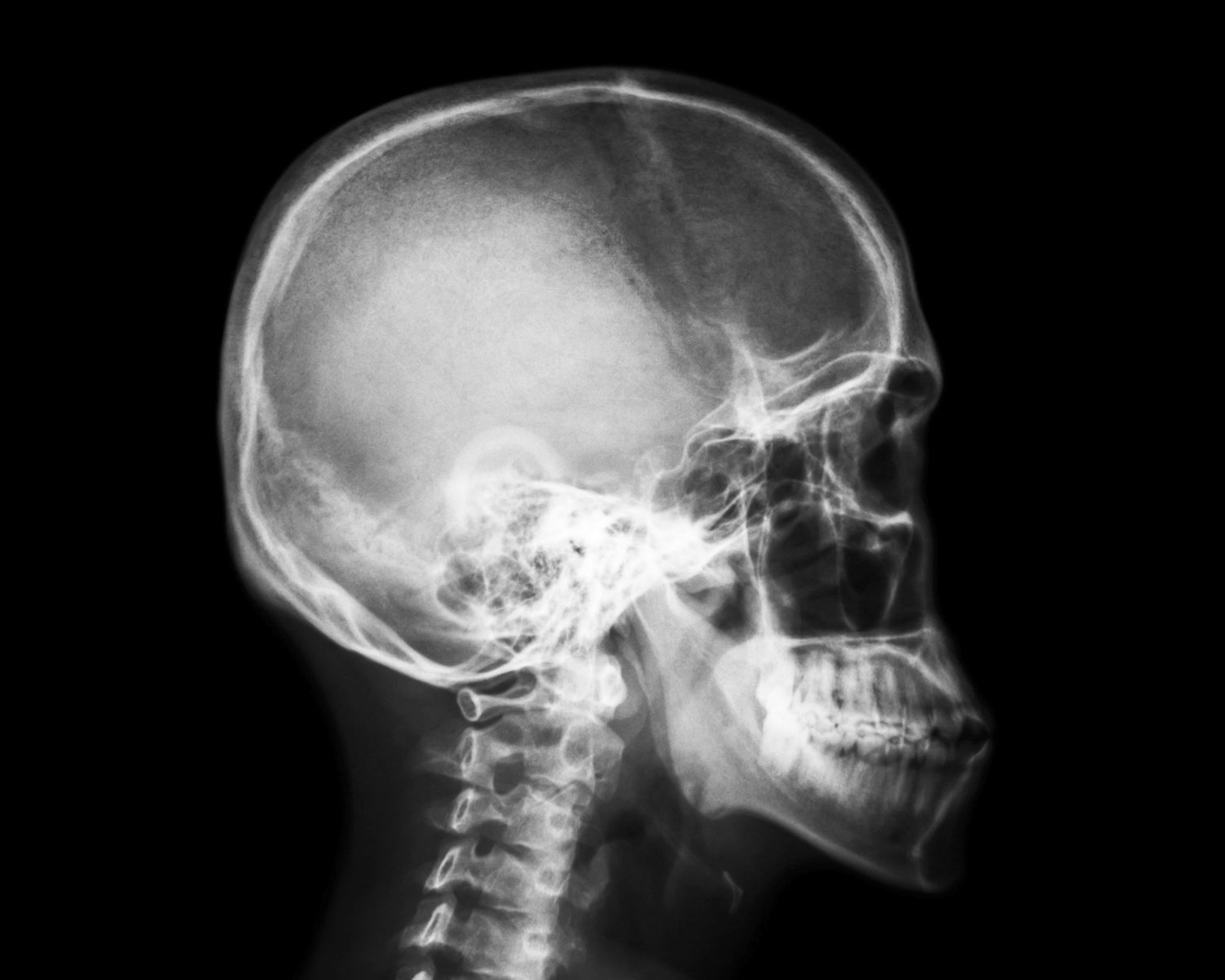
X-rays of the skull bones, commonly known as skull X-rays or cranial X-rays, may be performed for various medical reasons to assess and diagnose conditions related to the head and skull. Here are some common indications for getting a skull X-ray:
Head Trauma: After a head injury or trauma, a skull X-ray can be used to assess for fractures, signs of bleeding, or other structural damage to the skull bones.
Fractures: X-rays are useful in detecting fractures or breaks in the bones of the skull, which may occur due to accidents, falls, or other injuries.
Sinus Infections or Sinusitis: Skull X-rays may be used to identify signs of sinus infections or sinusitis, as these conditions can affect the bones and structures in the skull.
Dental Issues: In some cases, skull X-rays are used to evaluate dental problems, such as impacted teeth, dental abscesses, or abnormalities in the jaw.
Orthodontic Planning: Dentists and orthodontists may use skull X-rays for orthodontic planning to assess the alignment of the jaw and teeth.
Developmental Anomalies: X-rays can help identify congenital or developmental anomalies in the skull bones, such as craniosynostosis (premature fusion of skull bones).
Evaluation of Tumors: Skull X-rays may be part of the diagnostic process to identify tumors or abnormal growths in the skull.
Temporal Mandibular Joint (TMJ) Issues: X-rays may be used to assess the TMJ for conditions like temporomandibular joint disorder (TMD) or other joint abnormalities affecting the jaw.
It’s important to note that while skull X-rays provide valuable information about the bones of the skull, they are not as detailed as other imaging modalities, such as CT scans or MRI, which can provide more information about soft tissues and structures within the head. The choice of imaging modality depends on the specific clinical situation and the information needed by the healthcare provider.
As with any medical procedure, it’s essential to follow the guidance of your healthcare provider and the radiology staff, and to discuss any concerns or questions you may have about the procedure.
The preparation for a spine X-ray is generally straightforward, and there are usually minimal requirements. However, it’s essential to follow any specific instructions given by your healthcare provider or the imaging facility. Here are some general guidelines:
Clothing: You may be asked to change into a hospital gown, as buttons, zippers, or other metal components in regular clothing can interfere with the X-ray images.
Remove Metal Objects: You’ll likely be asked to remove any metal objects or jewelry from the area being X-rayed, as metal can interfere with the quality of the images. This includes items such as belts, watches, necklaces, piercings, and even underwire bras.
Inform the Technologist: Make sure to inform the X-ray technologist if there is any chance you could be pregnant, as they may need to take additional precautions to minimize radiation exposure to the fetus.
Follow Instructions for Positioning: The technologist will guide you into the appropriate positions for the X-ray. It’s important to follow their instructions to ensure accurate and clear images.
Stay Still during the Procedure: To obtain clear images, it’s crucial to remain still while the X-rays are being taken. The technologist may use immobilization devices or props to help you maintain the required positions.
Discuss Medical History: Inform the technologist and your healthcare provider about any relevant medical history, including prior surgeries or conditions that might affect the spine. This information helps ensure the imaging is tailored to your specific needs.
Generally, X-rays are safe and relatively quick procedures. The amount of radiation exposure is kept as low as reasonably achievable while still obtaining the necessary diagnostic information. If you have any concerns or questions about the procedure or its potential risks, don’t hesitate to discuss them with your healthcare provider or the radiology staff.
