03-543-6911

Ultrasound

Sound-based medical imaging technique.

Sonography of inguinal region

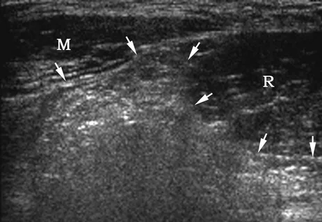
Sonography of the inguinal region, also known as inguinal ultrasound, is a diagnostic imaging procedure that uses ultrasound waves to examine the structures in the groin area. The inguinal region is located in the lower abdomen, near the groin, and it includes the inguinal canal, lymph nodes, blood vessels, muscles, and other tissues in that area.

  1. Evaluation of hernias: An inguinal ultrasound is commonly used to assess and diagnose hernias. It can help determine the presence, location, and size of a hernia, which is a protrusion of an organ or tissue through a weak spot in the abdominal muscles.
  2. Assessment of groin pain or swelling: If you are experiencing pain or swelling in the groin area, an inguinal ultrasound can help identify the underlying cause. It can detect abnormalities such as enlarged lymph nodes, fluid collections, or other soft tissue abnormalities.
  3. Examination of the inguinal canal: The inguinal canal is a passage in the abdominal wall through which structures, such as blood vessels and nerves, pass into the groin area. An inguinal ultrasound can evaluate the inguinal canal for abnormalities or obstructions that may be causing discomfort or other symptoms.
  4. Monitoring of lymph nodes: Lymph nodes in the inguinal region may become enlarged due to various reasons, such as infection or inflammation. Inguinal ultrasound can assess the size, shape, and characteristics of the lymph nodes to help determine the cause of the enlargement.
  1. Clothing: Wear comfortable clothing that allows easy access to the front of your neck. It is best to wear a shirt or blouse with an open collar or a top that can be easily removed.
  2. Medications: Continue taking your regular medications unless instructed otherwise by your healthcare provider.
  3. Fasting: In most cases, fasting is not necessary for a thyroid ultrasound. You can eat and drink normally.
  4. Inform the doctor: about any previous thyroid conditions, surgeries, or concerns.

During the procedure, you will be asked to lie on your back with a pillow or support under your shoulders to extend your neck. The technologist will apply a gel to the front of your neck and use a handheld device called a transducer to capture images of the thyroid gland. The transducer emits sound waves that create real-time images on a monitor.

After the procedure, the images will be reviewed by a radiologist who will interpret the findings and generate a report. Your healthcare provider will discuss the results with you and recommend any further actions if necessary.

It’s important to note that specific preparation instructions may vary depending on the healthcare facility and the reason for the thyroid ultrasound. Therefore, it is advisable to consult with your healthcare provider or the facility performing the ultrasound for any specific instructions or requirements.

فحص السونار على الغدة هو إجراء

Get Directions

Emergency Service?

Contact Us