03-543-6911

Ultrasound

Sound-based medical imaging technique.

Testicular & Scrotal ultrasound

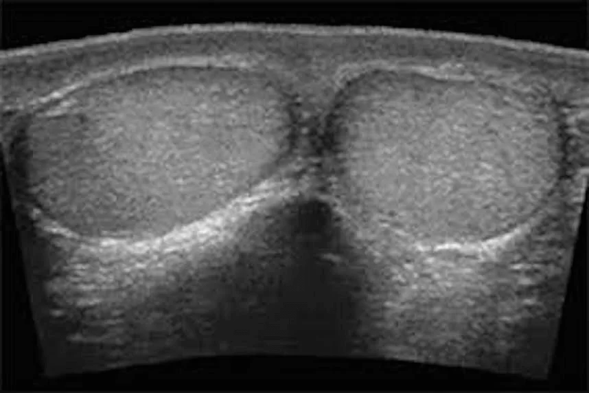
Testicular and scrotal ultrasound is a medical imaging procedure that uses ultrasound waves to examine the testicles and scrotum. It is a non-invasive and painless test that provides detailed images of the structures in this area. The testicles are the male reproductive organs responsible for producing sperm and testosterone, while the scrotum is the sac that holds the testicles.

  1. Evaluation of testicular pain or swelling: If you are experiencing testicular pain, swelling, or any abnormalities in the scrotal area, a testicular and scrotal ultrasound can help determine the cause. It can detect conditions such as testicular torsion (twisting of the testicle), epididymitis (inflammation of the epididymis), testicular tumors, hydrocele (fluid accumulation in the scrotum), or varicocele (enlarged veins in the scrotum).
  2. Assessment of testicular masses or lumps: If a lump or mass is found during a physical examination, a testicular and scrotal ultrasound can provide more information about its characteristics, size, and location. This helps in determining whether the mass is benign (non-cancerous) or requires further evaluation.
  3. Diagnosis of infertility: Testicular ultrasound can assess the size, shape, and blood flow to the testicles, which can help in evaluating male infertility. It can identify conditions such as testicular atrophy (shrinkage), varicocele, or other abnormalities that may affect sperm production and quality.
  4. Monitoring of testicular conditions: If you have a known testicular condition, such as a previous testicular injury or surgery, a testicular and scrotal ultrasound can be used to monitor the condition over time and assess any changes or progression.
  1. Clothing: Wear loose and comfortable clothing that allows easy access to the neck area. You may be asked to remove any jewelry or accessories around the neck.
  2. Fasting: In most cases, no fasting or dietary restrictions are necessary before a neck ultrasound. You can eat and drink normally.
  3. Medications: Continue taking your regular medications unless instructed otherwise by your healthcare provider.
  4. Inform the doctor: about any previous neck surgeries, medical conditions, or concerns you may have.

During the procedure, you will be asked to lie on your back on an examination table. The technologist will apply a gel to the neck area and use a handheld device called a transducer to move over the skin. The transducer emits sound waves that create images on a monitor, which the technologist will interpret and document.

After the examination, the images will be reviewed by a radiologist, who will generate a report. Your healthcare provider will discuss the results with you and recommend any further actions based on the findings.

Remember to consult with your healthcare provider for specific instructions and any additional preparations you need to undertake before a neck ultrasound.

Get Directions

Emergency Service?

Contact Us