- Clothing: Wear comfortable clothing that allows easy access to the front of your neck. It is best to wear a shirt or blouse with an open collar or a top that can be easily removed.
- Medications: Continue taking your regular medications unless instructed otherwise by your healthcare provider.
- Fasting: In most cases, fasting is not necessary for a thyroid ultrasound. You can eat and drink normally.
- Inform the doctor: about any previous thyroid conditions, surgeries, or concerns.
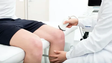
During the procedure, you will be asked to lie on your back with a pillow or support under your shoulders to extend your neck. The technologist will apply a gel to the front of your neck and use a handheld device called a transducer to capture images of the thyroid gland. The transducer emits sound waves that create real-time images on a monitor.
After the procedure, the images will be reviewed by a radiologist who will interpret the findings and generate a report. Your healthcare provider will discuss the results with you and recommend any further actions if necessary.
It’s important to note that specific preparation instructions may vary depending on the healthcare facility and the reason for the thyroid ultrasound. Therefore, it is advisable to consult with your healthcare provider or the facility performing the ultrasound for any specific instructions or requirements.
فحص السونار على الغدة هو إجراء